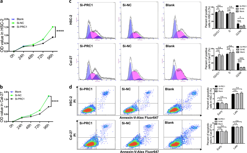
Fig. 3. Roles of PRC1 knockdown in proliferation, cell cycle and apoptosis in vitro.
a, b In the proliferation assay, the difference between si-PRC1-treated cells and control cells (si-NC and blank) was statistically significant after 48 h for Cal-27 and HSC-2 cells (pCal-27 = 0.003 and pHSC-2 = 0.000). c HSC-2 and Cal-27 cells in the si-PRC1-treated group exhibited higher G2/M phase arrest after 72 h compared to controls. d No statistically significant difference in cell apoptosis was observed in either early or late stages 72 h after transfection. All n = 3; error bars, mean ± SD; n.s, not significant, *p < 0.05, **p < 0.01, ***p < 0.001, ****p < 0.0001; t-test

