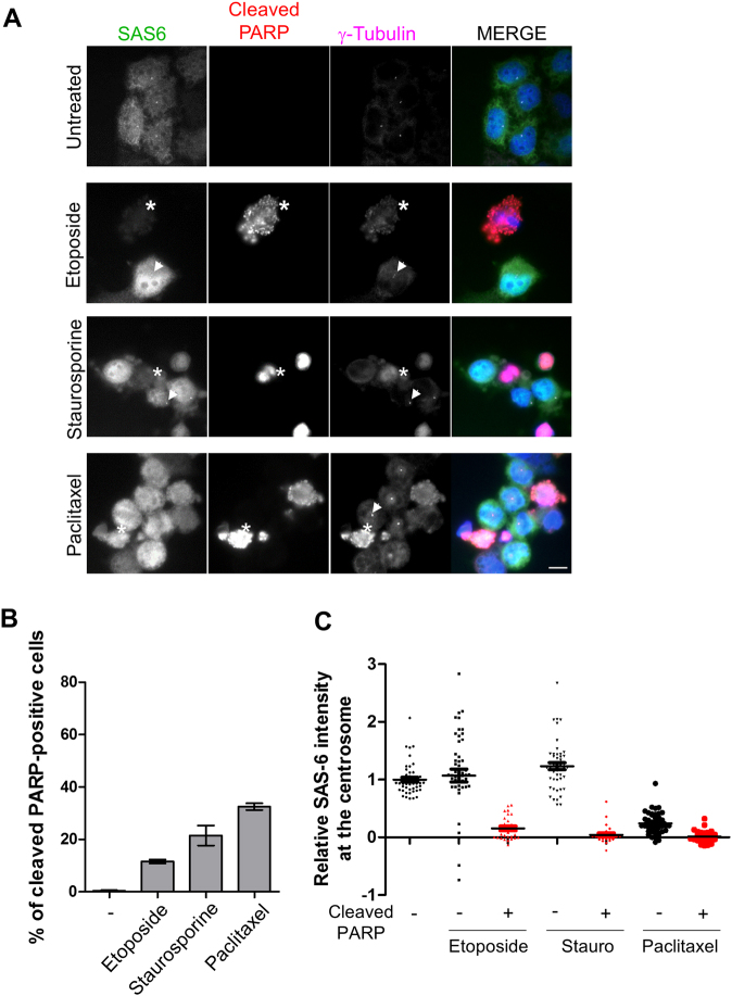
Fig. 5. Reduction of the centriolar SAS-6 levels in cells undergoing apoptosis.
a HeLa cells were treated with etoposide, staurosporine, or paclitaxel for 24 h and subjected to coimmunostaining with antibodies specific to SAS-6 (green), cleaved PARP-1 (red), and γ-tubulin (magenta). Asterisks indicate the cleaved PARP-positive cells whose SAS-6 intensities were significantly reduced. The arrowheads represent the cleaved PARP-negative cells with the remaining SAS-6 signals at the centrosomes. b Proportions of the cleaved PARP-1-positive cells were determined. c The centriolar intensities of SAS-6 were measured in cleaved PARP-1-negative or PARP-1-positive cells and analyzed with a scatter plot. Greater than 100 centrosomes per experimental group were analyzed in two independent experiments. *P < 0.05

