
Fig. 6. Effects of IDN-6556 on the centriolar SAS-6 levels in cells undergoing apoptosis.
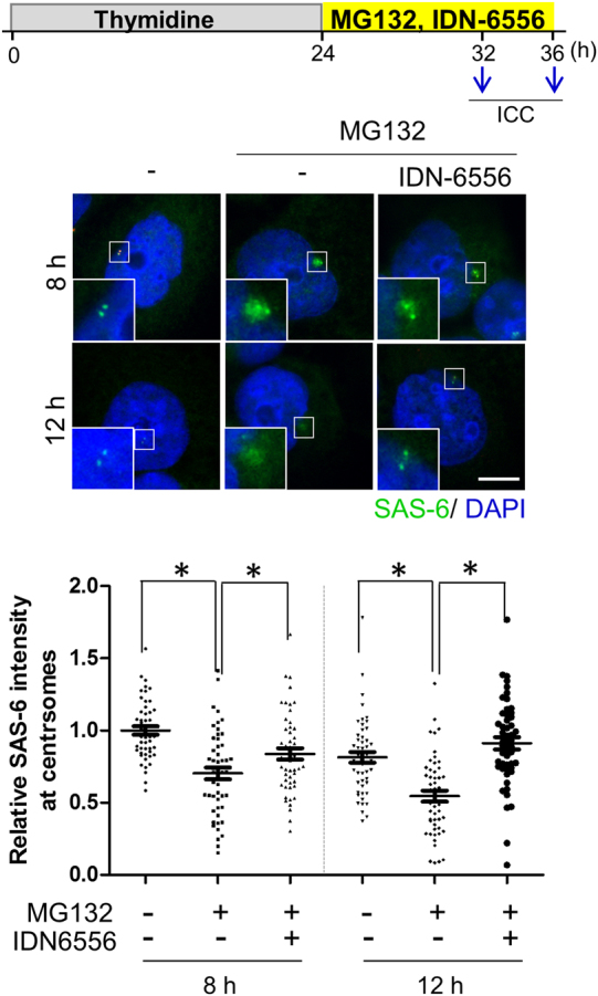
HeLa cells were treated with thymidine for 24 h, released into a fresh medium with MG132 and IDN-6566, and cultured for 8 or 12 h. The cells were immunostained with the SAS-6 (green) antibody. DNA was visualized with DAPI (blue). Scale bar, 10 μm. The centriolar intensity of SAS-6 was measured and analyzed with a scatter plot. Greater than 100 centrosomes per experimental group were analyzed in two independent experiments. *P < 0.05