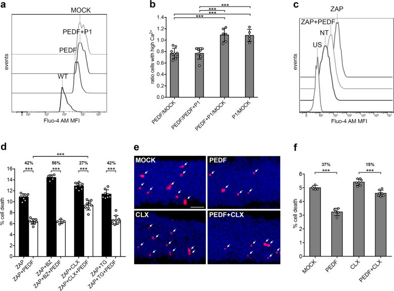
Fig. 1. Decreased intracellular Ca2+ after treatment with PEDF.
a Cytofluorimetric analysis of Ca2+, as evaluated by Fluo-4 AM labeling, in rd1 mutant photoreceptors not treated (MOCK) or treated with 6 pmol of PEDF (PEDF) or co-treated with PEDF and 10 molar excess of P1 peptide (PEDF + P1). As control, we show data from wild-type photoreceptors (WT). The levels of calcium (medium fluorescence intensity, MFI) in cells (events) were reduced in the presence of PEDF (PEDF vs MOCK and PEDF vs PEDF + P1). b Ratio of photoreceptor cells with high levels of Ca2+ (see parameters in Supplemental figure S1), as evaluated by Fluo-4 AM labeling and cytofluorimetric analysis, in PEDF treated compared with contralateral eyes treated either with vehicle (MOCK) or with PEDF and 10 molar excess of P1 peptide (PEDF + P1). Data are shown as means ± SD (N = 4–9; ***P ≤ 0.001). c Cytofluorimetric analysis of Ca2+, as evaluated by Fluo-4 AM labeling, in 661W cells stressed with 500 μM zaprinast (ZAP), to block PDE6 function and to mimic the rd1 mutation, and in 661W cells treated with Zaprinast and PEDF (ZAP + PEDF). As control, we show data from cells unstained with Fluo-4 AM (US) or not treated with zaprinast (NT). The levels of calcium (medium fluorescence intensity, MFI) in cells (events) were reduced in the presence of PEDF in cells stressed with zaprinast (ZAP + PEDF vs ZAP). d 661W cells were stressed with 500 μM zaprinast (ZAP) that caused cell death, as assessed by TUNEL staining. PEDF neuroprotective effects were maintained after exposure to 100 μM 3′,4′-dichlorobenzamyl (BZ) or 200 nM Thapsigargin (TG). PEDF neuroprotection was reduced when cells were exposed to 10 μM caloxin (CLX). The percentage of PEDF neuroprotection is reported above the P-value. Data are shown as means ± SD (N = 5–8; *** P ≤ 0.001). e Sections of rd1 mutant retinas either treated with vehicle (MOCK) or with 6 pmol of PEDF (PEDF) or with 100 μM of CLX or with PEDF and CLX (PEDF + CLX). The outer nuclear layer of the retina, containing the photoreceptor nuclei (blue), is shown after TUNEL staining (red, arrows). Scale bar: 20 μm. f Quantification of cell death in rd1 mutant retinas either treated with vehicle (MOCK) or with 6 pmol of PEDF (PEDF) or with 100 μM of CLX or with PEDF and CLX (PEDF + CLX). The percentage of neuroprotection is reported above the P-value. Data are shown as means ± SD (N = 4–6; ***P ≤ 0.001)

