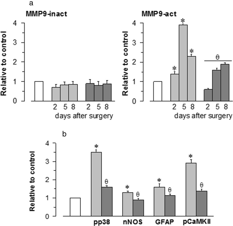
Fig. 5.
Stimulation of MMP-9 protein expression and inflammatory markers via pMCAO. a The expression levels of both the active (86 kDa) and the inactive (92 kDa) forms of MMP-9 were determined at different times after surgery. b The ischaemic-induced phosphorylation of p38 and CaMKII as well as the expression levels of nNOS and GFAP were evaluated 8 days after surgery. The enhancing effects of pMCAO (light grey) were reduced by S1RA administration (dark grey). Each bar represents the means ± SEM of the data from three determinations performed using different gels and blots. Groups of 8–10 mice were used for each interval. Immunosignals (average optical density of the pixels within the object area/mm2; Quantity One Software, Bio-Rad, Madrid, Spain) were expressed as the change relative to the sham-operated group (attributed an arbitrary value of 1; white bars). Asterisk denotes the significant difference from the sham-operated mice, θ from the pMCAO group, all pairwise Holm-Sidak multiple comparison test following ANOVA, p < 0.05

