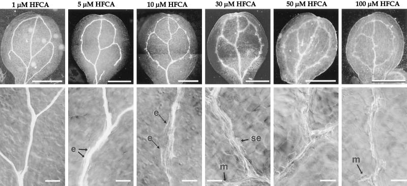
Figure 4.
Vein pattern of cotyledon from auxin polar transport inhibitor-grown seedlings. The top row are dark-field images of Ler cotyledons from plants grown in the presence of the polar auxin transport inhibitor HFCA. The bottom row contains differential interference contrast images of the midvein between or adjacent to the branch site of the distal secondary. Arrows with an “e” point to short files of ectopic TEs, arrows with “se” point to solitary ectopic tracheary elements, and arrows with an “m” point to ectopic misshapen tracheary elements. Size bars for the top row = 1 mm; for the bottom row = 100 μm.

