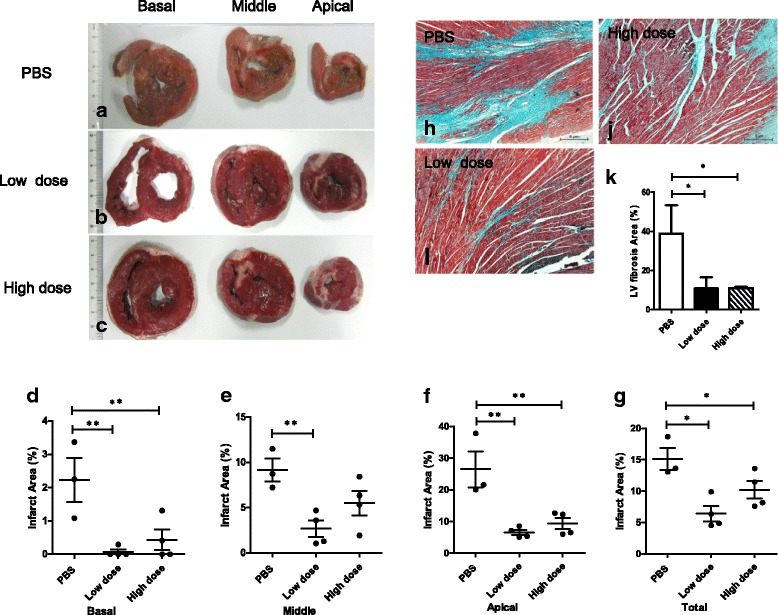
Fig. 5.

Pathological findings showing that intravenous injection of allogeneic UC-MSCs reduced the infarct area of the left ventricular myocardium at week 8 after AMI. a–c Representative histological images of left ventricular basal, middle, and apex cross-sections after TTC staining in the three groups. d–g Infarct area measured by TTC staining was lower in the UC-MSC-treated animals after 8 weeks. h–j Representative photomicrographs of the left ventricle (LV) highlight the collagen deposition (blue) on the infarct region of the three groups by Masson’s trichrome staining. Scale bars = 5 μm. k Quantitative summary of LV infarct area by Masson’s trichrome staining. Data are presented as the mean ± SD. Phosphate-buffered saline (PBS) group, n = 3; low-dose group, n = 4; and high-dose group, n = 4. *P < 0.05, **P < 0.01
