
Fig. 2.
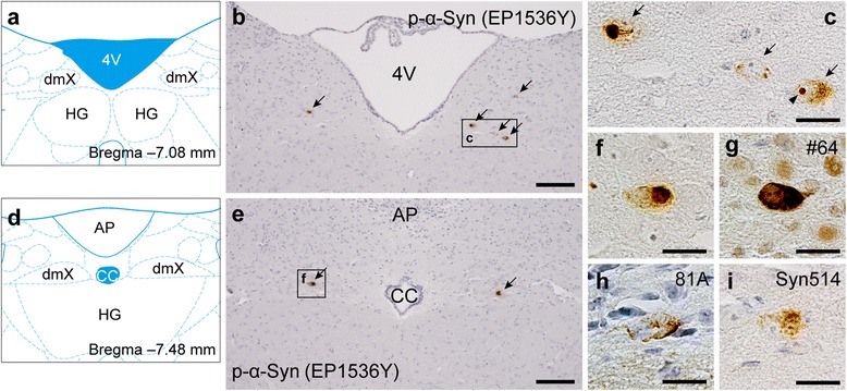
Phosphorylated α-synuclein (p-α-Syn) pathology in the dmX 45 days after inoculation of α-Syn preformed fibrils. a Schematic of anatomy at bregma − 7.08 mm modified from [47]. dmX, dorsal motor nucleus of the vagus nerve; HG, hypoglossal nucleus; 4V, fourth ventricle. b P-α-Syn (EP1536Y) immunohistochemistry around bregma − 7.08 mm. Scale bar 100 μm. c High-magnification image of p-α-Syn–positive cells (arrows). P-α-Syn–positive intranuclear dots are occasionally observed (arrowhead). Scale bar 20 μm. d Schematic of anatomy at bregma − 7.48 mm. AP, area postrema; cc, central canal. e P-α-Syn (EP1536Y) immunohistochemistry around bregma − 7.48 mm. Scale bar 100 μm. f High-magnification image of p-α-Syn–positive cells. Scale bar 20 μm. g–i Immunohistochemistry assessing p-α-Syn (#64), p-α-Syn (81A), or nitrated α-Syn (Syn514) shows p-α-Syn–positive or nitrated α-Syn–positive cells in the dmX. Scale bar 20 μm