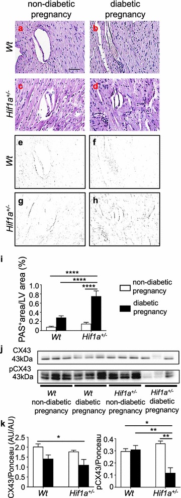
Fig. 6.

The amount of AGEs is increased whereas the activity of gap junction CX43 is reduced in the heart of diabetes-exposed Hif1a+/− offspring. Representative images of PAS staining of advanced glycation end products (AGE; a–d). Scale bar = 50 µm. e–h Delineated PAS+ area in the myocardium by Adobe Photoshop. i Quantification of PAS staining determined as a percentage of positive area in the field of view by ImageJ. The values are mean ± SEM (n = 4). Representative Western blots and quantification of both CX43 and its phosphorylated isoform pCX43 in the LV myocardium of the offspring (j, k). The values are mean ± SEM (n = 3). Statistical significance assessed by two-way ANOVA: diabetes effect (CX43, P = 0.0088) and interaction between genotype and diabetes (AGEs, P = 0.0013; pCX43, P = 0.0030) followed by post hoc Tukey’s multiple-comparison test, *P < 0.05, **P < 0.01, ****P < 0.0001. CX43 connexin 43, PAS periodic acid-shiff, AU arbitrary units
