Fig. 8.

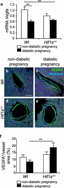
Changes in the expression of Vegfa in the heart of the diabetes-exposed offspring. Relative mRNA levels of Vegfa in the LV myocardium of the heart of 12-week-old offspring by RT-qPCR (a), the values are mean ± SEM (n = 8). Data are normalized to Hprt1 mRNA of control gene. b–e Representative confocal images of immunohistochemical staining of VEGFA (green) in the wall of coronary vessels in the LV myocardium of the 12-week-old offspring, Hoechst stained cell nuclei (blue). Scale bar = 50 µm. f A relative quantification of VEGFA in the blood vessels is determined as a percentage of VEGFA+ area per blood vessel area. The values are mean ± SEM (n = 4). Statistical significance assessed by two-way ANOVA: diabetes effect (Vegfa mRNA, P = 0.0003) and interaction between genotype and diabetes (VEGFA in blood vessels, P = 0.0382) followed by post hoc Tukey’s multiple-comparison test, *P < 0.05, **P < 0.01
