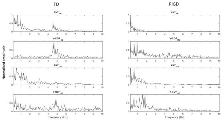
Figure 1.
Power spectrum of COP and COP velocity of a tremor dominant (TD) patient and a postural instability and gait difficulty (PIGD) patient for both the medial–lateral (ML) and anterior–posterior (AP) directions. The graphs on the left and right sides of the page present the power spectrum signal of a TD patient and a PIGD patient, respectively. COPML: COP in the ML direction, COPAP: COP in the AP direction, V-COPML: COP velocity in the ML direction, and V-COPAP: COP velocity in the AP direction.

