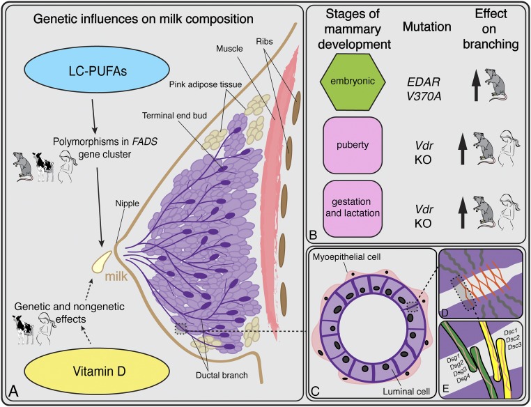
Fig. 2.
Overview of breast anatomy, development, milk production, and histology. (A) Genetic influences on milk production summarized from the main text. The rodent, human, and cow figures indicate the systems from which the results are known. (B) Three stages of mammary development alongside the genetic mutations known to increase ductal branching. Hormones induce the pubertal and gestation/lactation stages (denoted in pink). In vitamin D-deficient adipose tissue, ductal branching increases during these two stages of development. The embryonic stage (in green) is not hormonally induced; branching density is influenced by the ectodysplasin pathway (108) and, specifically, increased by EDAR V370A. (C) Cross-section of a ductal lumen showing the bilayer of luminal and myoepithelial cells. (D) Schematic of a desmosome, one of the main adhesive structures between mammary luminal cells. The orange lines represent desmosomal cadherins in the extracellular space. The gray lines in the purple area represent the intracellular filaments. (E) Close-up view of the desmocolin proteins that comprise the desmo-adhesome. An increase in the activity of the ectodyplasin pathway alters the relative proportions of Dsc2 and Dsc3, leading to a reduction in the adhesive strength.

