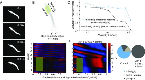
Fig. 3.
Midbody B-type motor neurons generate rhythmic activity independent of proprioceptive coupling. (A) Time-lapse video images from a recording when B-type motor neurons in an anterior body region (10–30% along the worm body) were optogenetically inhibited. (B, Upper) Schematic illustrates the effect of spatially selective inhibition of B-type motor neurons. Optogenetic inhibition of anterior B-type motor neurons induced high-frequency undulation in the posterior region. (Lower) Representative curvature kymograph. Green shaded region shows the selected spatiotemporal region for optogenetic inhibition. (C) C. elegans undulation frequency at different viscosity. Black line is undulation frequency of control animals; blue line is midbody undulation frequency when anterior bending activity was abolished. Error bars are SEM; n ≥ 8 worms for each data point. (D) Representative curvature kymograph during optogenetic inhibition of anterior B-type motor neurons, with and without midbody B-type neurons (DB 4–5 and VB 5–7). (E) Pie chart summarizes the percentage of locomotor states when anterior bending activity was abolished. h-f wiggle: midbody undulation frequency was higher than that before anterior bending activity was abolished; non–h-f wiggle: midbody undulation frequency was equal to or less than that before anterior bending activity was abolished; paralysis: no waves emerged in the midbody. Control (Pacr-5::Arch), n = 241 measurements, 20 worms; midbody B-type neuron-ablated worms (Pacr-5::Arch; Pacr-5::miniSOG), n = 77 measurements, 11 worms.

