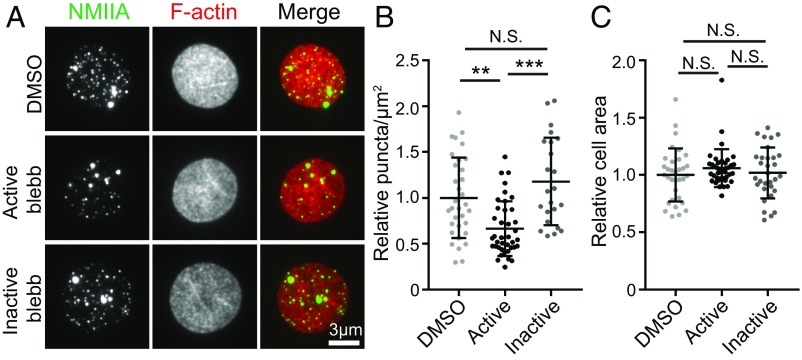
Fig. 3.
NMIIA motor activity controls the association of NMIIA puncta with the RBC membrane. (A) TIRF microscopy of NMIIA motor domain (green) and rhodamine-phalloidin for F-actin (red) in human RBCs pretreated with DMSO alone, 20 μM active blebbistatin (blebb), or 20 μM inactive blebb before fixation and immunostaining. RBCs were flattened by centrifugation onto glass coverslips before imaging to visualize a larger area of the membrane. (B) NMIIA puncta density at the RBC membrane measured as the number of NMIIA puncta per square micrometer in TIRF images. DMSO versus active blebbistatin (**P = 0.0016) and active blebbistatin versus inactive blebbistatin (***P < 0.0001) are shown. (C) Cell surface areas for RBCs in TIRF images in each treatment group. There is no significant difference in cell areas between treatment groups by one-way ANOVA. Cells from two individual donors are shown: DMSO (n = 33), active blebbistatin (n = 40), and inactive blebbistatin (n = 31). N.S., not significant.

