Fig. 1.
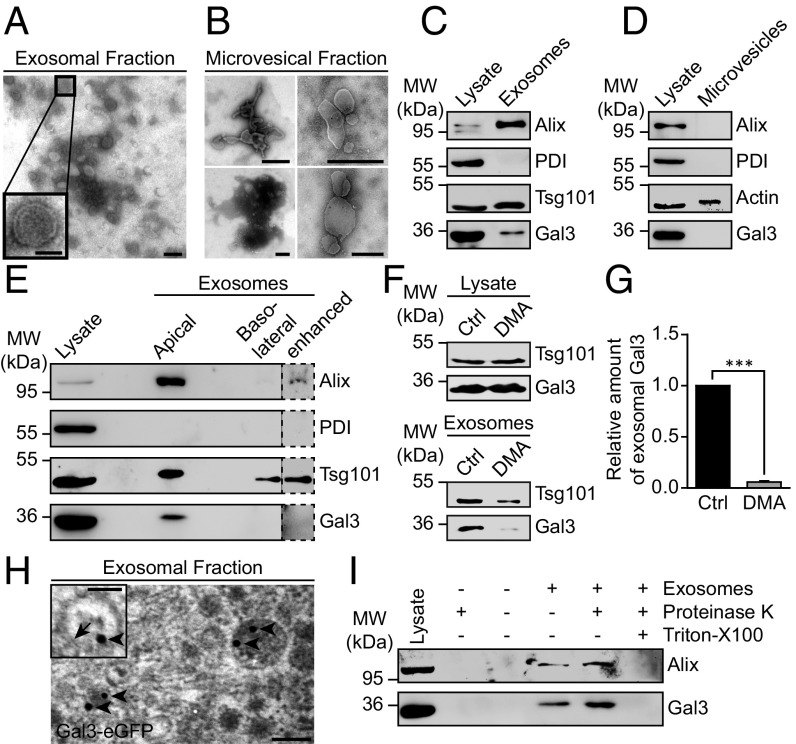
Gal3 is contained in exosomes. (A and B) The 100,000 × g pellets (exosomes) (A) or the 10,000 × g pellets (microvesicles) (B) were subjected to negative staining for electron microscopy. Inset shows the typical appearance of an exosome. (C) Western blotting analysis of the exosomal fraction. Representative results, n = 3 independent experiments. (D) Western blotting analysis of the microvesicular fraction. Actin served as loading control for microvesicles. Representative results, n = 3 independent experiments. (E) Immunoblot analysis of the exosomal fractions from filter-grown MDCK cells. Representative results, n = 3 independent experiments. (F) Western blotting analysis of DMA-treated cells. (G) Quantification of experiments as in F. Normalized to the respective cell lysate. Means ± SEM, n = 5 independent experiments. (H) Electron microscopy analysis of Gal3-eGFP localization in exosomes. A total of 15 nm gold-labeled GFP nanobodies (arrowheads) was detected in exosomes, often in close proximity to the exosomal membrane (arrow). (I) Proteinase protection assay. Representative results, n = 3 independent experiments. Statistical analysis: Student’s unpaired t test, ***P < 0.001. [Scale bars: A and H, 100 nm; A and H, (Inset), 50 nm; B, 500 nm.]

