Fig. 4.
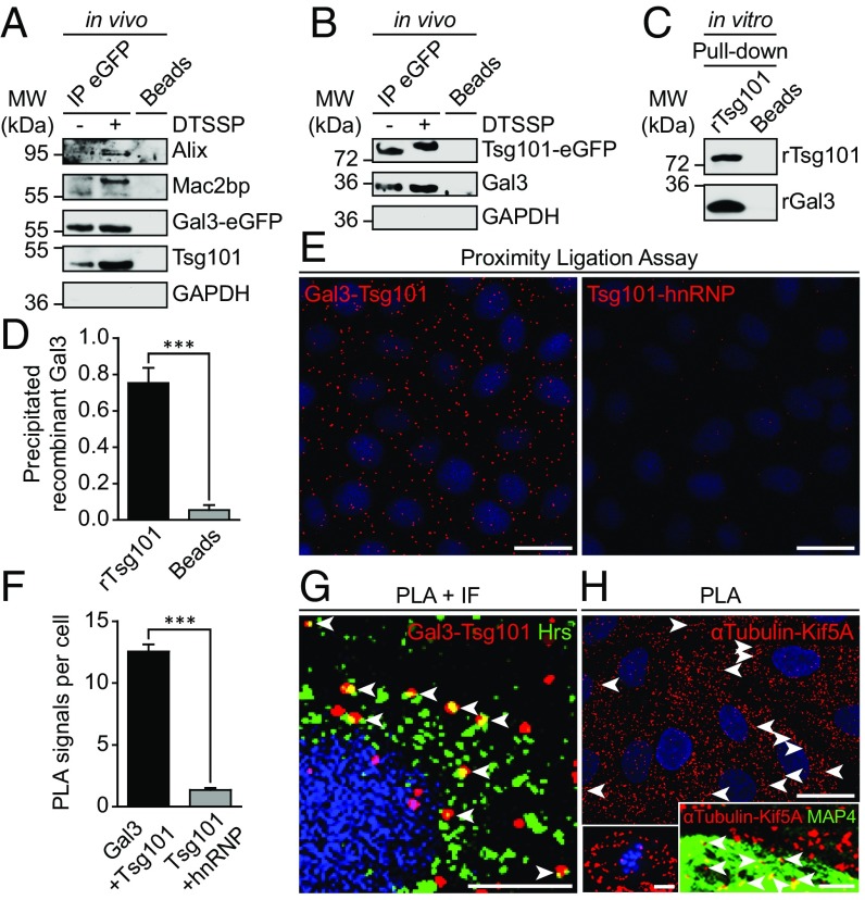
Gal3 interacts with the ESCRT-component Tsg101. (A) Coimmunoprecipitation of Gal3-eGFP in MDCK cells. Gal3-eGFP fusion protein and its binding partners were immunoprecipitated with GFP-nanobody beads and detected by immunoblotting. Positive control: Alix, Mac2bp; negative control: GAPDH. Representative results, n = 3 independent experiments. (B) Complementary approach with Tsg101-eGFP. Negative control: GAPDH. Representative results, n = 3 independent experiments. (C) Direct rGal3 and rTsg101-GST interaction as determined by in vitro pull-down assay (rGal3: recombinant Gal3; rTsg101: recombinant Tsg101). Representative results, n = 4 independent experiments. (D) Quantification of experiments as in C. Means ± SEM, n = 4 independent experiments. (E) Physical closeness of the two proteins was evaluated by an in situ proximity ligation assay (PLA). Negative control: hnRNP. (F) Quantification of experiments as in E. Means ± SEM, 10–15 cells per experiment, n = 3 independent experiments. (G) PLA and immunofluorescence (IF) were combined to ensure that the interaction between Gal3 and Tsg101 was localized to Hrs-positive vesicles (arrowheads). (H) PLA positive control with α-tubulin and Kif5A. Single microtubules were decorated PLA signals (arrowheads). (Left Inset) Mitotic cell in metaphase with PLA signals visualizing microtubule-Kif5A proximity on the mitotic spindle apparatus. (Right Inset) Combination of PLA with MAP4 staining (arrowheads; see also SI Appendix, Fig. S6A). Statistical analysis: Student’s unpaired t test, ***P < 0.001. [Scale bars: E and H, 25 µm; G, 10 µm; H (Insets), 5 µm.]

