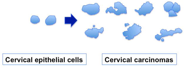
Figure 2. Nuclear shape changes in cervical cancer.
Outlines of nuclei were traced from images of sections of normal cervical epithelium and representative cervical carcinomas. Most nuclei in carcinoma specimens are larger and exhibit various irregular shapes. Additionally, micronuclei and nuclear fragments often associate with the cancer nuclei.

