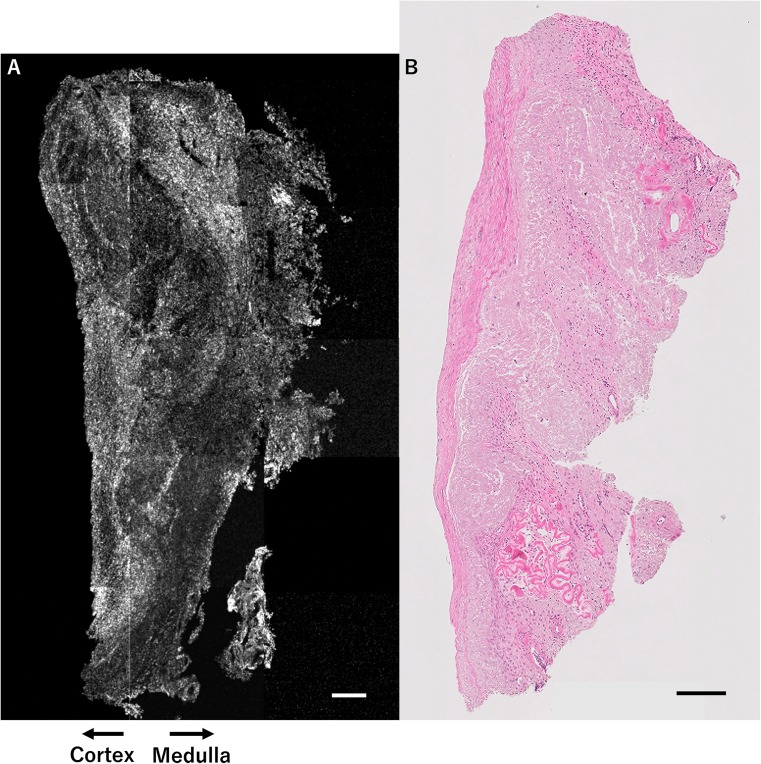
Fig. 2.
OCT image and H&E-stained histological image of ovarian tissue from patient 2 (menopausal case). Results of OCT examination of ovarian tissue from patient 2, who was 45 years of age with severely diminished ovarian reserve. No follicles are detected by OCT (a), in accordance with histological examination (b). This OCT image reflects the poor result of the ovarian reserve test (FSH 20.1 mIU/ml, AMH 0.51 ng/ml at 1.5 years prior to ovariectomy). Scale bar = 200 μm (a, b). OCT optical coherence tomography, H&E hematoxylin and eosin, FSH follicle-stimulating hormone, AMH anti-Müllerian hormone

