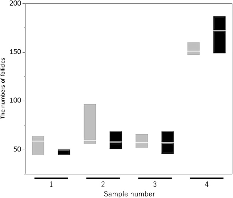
Fig. 5.
A comparison of follicle counts between OCT and histological examination. There is no significant difference in follicle counts between OCT and histological examination (P = 0.78, Wilcoxon signed-rank test). Sample number 1–3: patient 3, 4: patient 4. Gray boxplot: OCT examination, Black boxplot: histological study

