
Fig. 2.
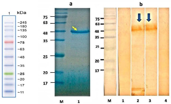
SDS-PAGE analysis of purified antigen and Western blot analysis of rDgK antigen. (a) Purified protein was loaded on 12% acrylamide gel, stained with Coomassie brilliant blue R-250. Lane M, protein molecular standard; lane 1, SDS-PAGE of purified recombinant protein (arrow). (b) Lane M, protein molecular standard; lane 1, Western blotting of expressed rDgK probed with (lane 1) the negative control pool sera, (lane 2) suspected dog pool sera, and (lane 3) the positive-control serum pools (arrows). Lane 4 indicates total protein of untransformed E. coli BL21 in Western blot with the positive-control serum pools.