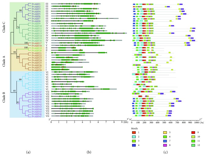
Figure 3.
Exon/intron structure of PvARF genes. (a) Phylogenetic tree of PvARF proteins constructed using MEGA5 based on the multiple alignments of full-length amino acids. (b) Exon/intron arrangements of PvARF genes. Exons and introns are represented by green boxes (open reading frame in green, untranslated region (UTR) in gray), and black lines, respectively, and their sizes are indicated by the scale at the bottom. The red vertical bar denotes the targets of Osa-miR167a in PvARF genes; the red arrows denotes the targets of Osa-miR160a in PvARF genes. (c) Schematic representation of conserved motifs in the PvARF proteins predicted by MEME. Each motif is represented by a number in the colored box. The black lines represent nonconserved sequences. Lengths of motifs for each PvARF protein were exhibited proportionally.

