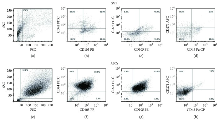
Figure 1.
Flow cytometry analysis of mesenchymal cells in freshly isolated and cultured SVF. Dot plots show the morphology of SVF (a) and ASCs (e). SVF is a heterogeneous cell population, containing CD105-, CD44-, CD73-, and CD271-positive mesenchymal cells (b, c) and a small fraction of CD45-positive cells (d), due to the normal presence of leukocytes in SVF. After 15 days of culture, a large and enriched population of ASCs highly expresses mesenchymal markers (f, g), whereas it is completely negative for CD45.

