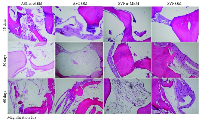
Figure 3.
H&E staining to monitor SB colonization by ASCs and SVF. Both ASCs (left panels) and SVF (right panels) grow on SB in the absence of α-MEM or in osteogenic medium (OM). The presence of new tissue formation is evident since 15 days of culture, and it increases over time. Images of H&E staining are reported for each time point (15, 30, and 60 days). Magnification 20x.

