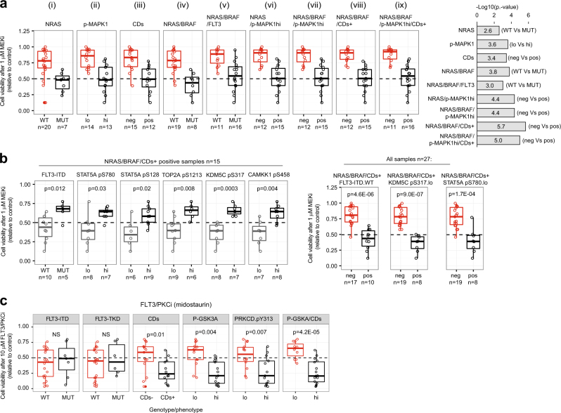
Fig. 2.
Integration of genomic, phosphoproteomics, and mass cytometry data to rationalize kinase inhibitors sensitivity. a Viability of AML cells within the indicated genotype/phenotype groups after treatment with MEKi. b Sensitivity of NRAS/BRAF/CDs+ positive cells to MEKi as a function of the indicated factors. c FLT3/PKCi sensitivity of AML cells with the indicated phenotype/genotype. Phosphorylations are denoted as (hi) and (lo) based on a greater or lower phosphorylation than the median across all cases. Significance was assessed by Mann–Whitney test

