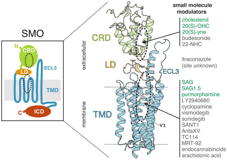
Figure 2.
The overall structure of SMO. SMO consists of a large extracellular region, made up of the CRD (green) and LD (orange), and an intracellular domain (ICD, red) in addition to the seven-pass α-helical transmembrane domain (TMD, blue) (left panel). The multi-domain SMO crystal structure revealed a stacked domain arrangement with two physically separable binding sites (right panel). The approximate location of the two binding sites is marked with dashed black ovals in both left and right panels. ECL3 and TMD helix VI are also labeled. Agonist (green) and antagonist (dark grey) small molecule modulators are listed on the right and associated with a particular binding site, if known. This list is not exhaustive.

