Table 1.
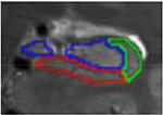
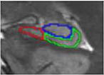
Slice types and features, based on La Joie et al., 2010.
| Slice type | Typical number of slices |
Defining feature |
Outter Boundaries | Internal boundaries (if novel or changed from previous slices) |

|
|---|---|---|---|---|---|
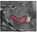
| a. Head – anterior, subiculum only | 1 to 2 | In head, but no SRLM | alveus superiorly (inclusive), parahippocampal WM inferiorly, CSF laterally | n/a |
|
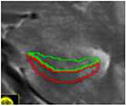
| b. Head – anterior, subiculum and CA1 | 1 to 2 | In head, SRLM | alveus superiorly (inclusive), parahippocampal WM inferiorly, CSF laterally | The subiculum was separated from CA1 by
the darker band (representing the stratum lacunosummoleculare - SRLM) and white voxels of CSF from uncal sulcus |
|
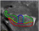
| c. Head – with digitations | 2 to 3 | In head, clear “digits” | alveus superiorly (inclusive), parahippocampal WM inferiorly, CSF laterally | CA1 was traced on both the medial and
lateral ends of the superior/ dorsal GM band, while the middle digitation (or medial digit when only 2 were present) of this superior GM band corresponded to the CA2-4/DG subfield. Vertical lines are traced to separate CA1 from other subfields on this slice |
|
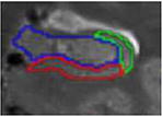
| d. Head – post digitations | 1 to 2 | In head, post digits | alveus superiorly (inclusive), parahippocampal WM inferiorly, CSF laterally | CA1 was traced on the lateral end of the
superior/dorsal GM band. In agreement with Joie et al., 2010 and Harding et al., 1998, the CA1/subiculum border was not constant along the medial–lateral axis: in the most anterior slices of the body of the hippocampus, most of the inferior part of the hippocampus was considered as subiculum, while in the most posterior slice, CA1 progressed medially |
|
| e. Head – uncal apes | 1 | Last slice in head, uncal apex visible | alveus superiorly (inclusive), parahippocampal WM inferiorly, CSF laterally | same as above |
|
| f. Body | 9 to 10 | post uncal apex | alveus superiorly (inclusive), parahippocampal WM inferiorly, CSF laterally, CSF medially | Vestigial hippocampal sulcus used as the infero-medial border of the CA2-4/DG subfield |

|
| g. Body – last slice | 1 | slice before fornix separates | alveus superiorly (inclusive), parahippocampal WM inferiorly, CSF laterally, CSF medially | CAl/subiculum border is placed from the
intersection between CA2-4/DG and subiculum subfields to the adjacent parahippocampal gyrus white matter |
|
