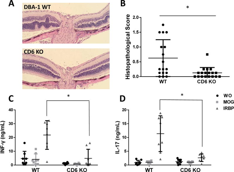
Figure 2.

Histopathological and immunological analyses of wild-type (WT) and CD6 knockout (KO) mice with experimental autoimmune uveitis (EAU). A and B: Representative histopathological images and scores for WT and CD6 KO on day 21 after immunization to induce EAU. The WT samples exhibited prominent retinal folds and occasional protrusions, whereas CD6 KO mice exhibited significantly fewer histopathological changes and lower scores. Data are shown as means ± standard deviations (SD). N=17 and 18 in the WT and CD6 KO groups, respectively. * p <0.05, Student’s t-test. C and D: CD6 KO mice exhibited decreased IRBP-specific T cell responses after immunization. Data are shown as means ± SD. N=8, one-way ANOVA.
