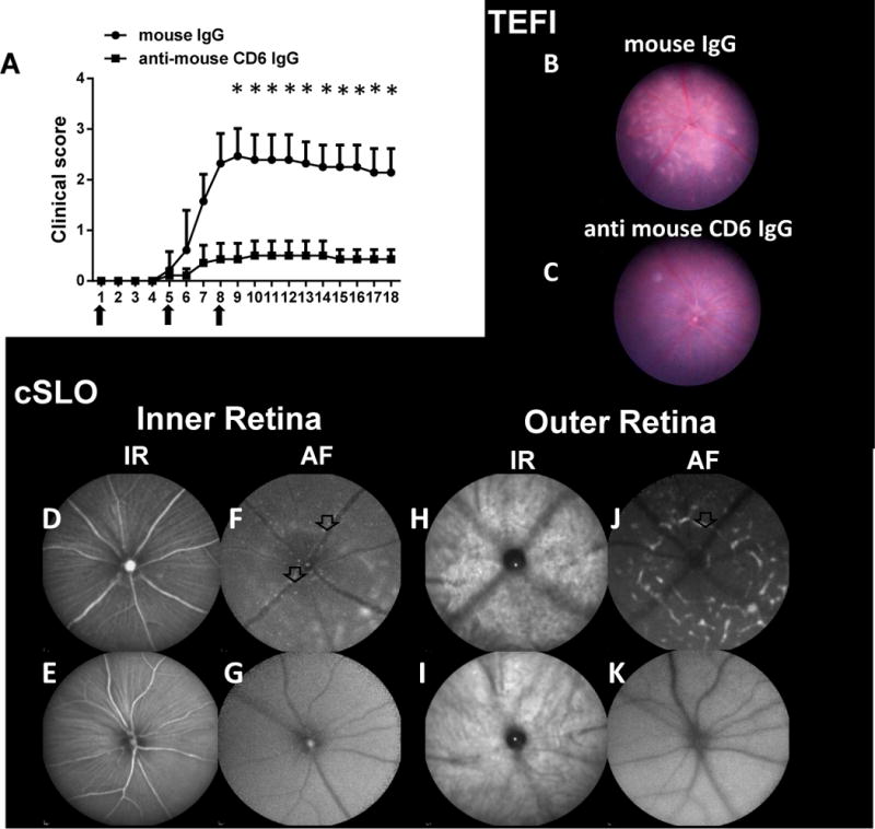
Figure 4.

Treatment studies using a newly developed mouse anti-mouse CD6 mAb. A: Mice with experimental autoimmune uveitis (EAU) induced by the adoptive transfer of uveitogenic T cells exhibited reduced clinical scores when treated with the mouse anti-mouse CD6 mAb clone 6C1. Briefly, 200 μg of 6C1 or mouse IgG were administered on days 0, 4 and 7 (arrows). Data are shown as means ± standard deviations . N=7 per group. * p <0.05, two-way ANOVA. B–K: Representative images from topical endoscopy fundus imaging (TEFI) and confocal scanning laser ophthalmoscopy (cSLO) of control and CD6 mAb-treated mice on Day 8 after adoptive transfer. B and C: TEFI images revealed reduced retinal damage caused by uveitogenic T cells after treatment with the anti-mouse CD6 IgG. Multiple retinal lesions (arrow) were observed in mice treated with IgG (control), whereas many fewer lesions were observed in CD6 mAb-treated mice. D–K: Hyper-reflective foci (arrows) and large retinal lesions (marked area) were observed in cSLO images from the control group, but were rarely observed in images from the CD6 mAb-treated group.
