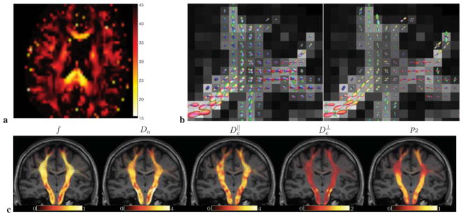
Figure 6. ODF dispersion, local kernel-based tractography, and parameter maps.
a, Fiber orientation dispersion angle θdisp from p2 (prevalence method) emphasizes major WM tracts. b, Empirical reference ODF ℘(0) (left, see text and Appendix G), and the fiber ODF calculated using plm from Eq. (3) (right), for the b = 5 shell, with l ≤ lmax = 6. Note the strong ODF sharpening effect, due to the deconvolution with locally estimated kernels Kl(b, x). c, Coronal view of a structural MR image (MPRAGE) overlaid with a reconstruction of the corticospinal fiber tract colored according to the value of the respective parametric maps x and p2 (via prevalence method, Fig. 3) at each point along the track. Fiber tracts have been reconstructed using improved probabilistic streamline tractography (Tournier et al., 2010) by integration over local fiber ODFs calculated using plm estimated from Eq. (3) using the voxel-wise estimated kernel Kl(b, x), for the b = 5 shell with maximal SH order lmax = 6.

