Figure 1.
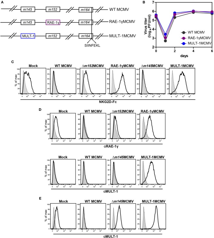
Recombinant viruses used in this study. (A) Recombinant murine CMV (MCMV) was made by insertion of genes for NKG2D ligands, RAE-1γ, and murine UL16 binding protein-like transcript-1 (MULT-1) in place of m152 and m145, respectively. OVA-derived Kb restricted CD8+ T cell epitope SIINFEKL was swapped with Dd restricted viral CD8+ T cell epitope of m164 167AGPPRYSRI175. (B) Multi-step growth kinetics assay on mouse embryonic fibroblasts (MEF) comparing wild-type (WT) MCMV, RAE-1γMCMV, and MULT-1MCMV is shown. (C,D) MEFs were infected with 1.5 PFU/cell of indicated viruses and expression of NKG2D ligands was evaluated 24 h after infection by staining either with (C) mouse NKG2D-Fc fusion protein (black line) or (D) αRAE-1γ (upper row, black line), αMULT-1 (lower row, black line), and appropriate isotype controls (gray). (E) SVEC4-10 cells were infected with 3 PFU/cell of WT MCMV, Δm145MCMV, and MULT-1MCMV for 16 h. Surface expression of MULT-1 was detected with αMULT-1 (black line) or isotype control (gray).

