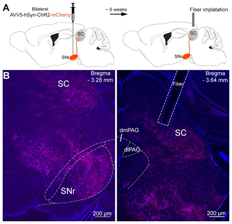
Figure 2.
Infusion of viral vectors into the SNr. (A) Viral vectors were injected into the SNr, resulting in the expression of either Channelrhodopsin-2 (ChR2)-mCherry (n = 10) or mCherry (n = 8) in the nigrotectal pathway. The optical fibers were placed above the SC. (B) Representative coronal photomicrographs showing the expression of ChR2-tdtomato in SNr somata, as well as in SNr terminals within the periaqueductal gray (PAG) matter and SC (blue: DAPI, red: tdtomato).

