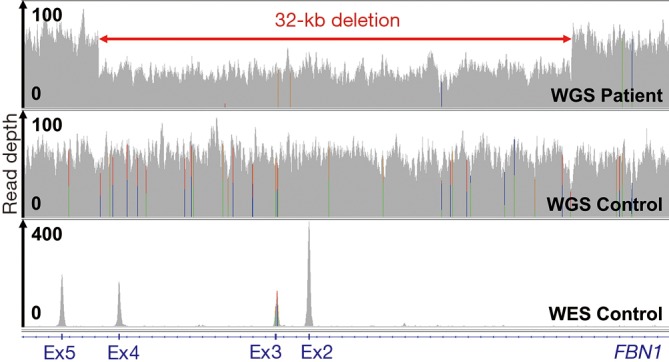
Figure 3.

CNV detection by NGS. Read depth (coverage tracks) of 60× PE150 PCR-free WGS data of the index patient and a control as well as 100× PE100 WES data (12) of a control for the deleted and flanking genomic regions displayed in IGV. CNV, copy number variation; NGS, next-generation sequencing; WES, whole-exome sequencing; WGS, whole-genome sequencing.
