Figure 9.
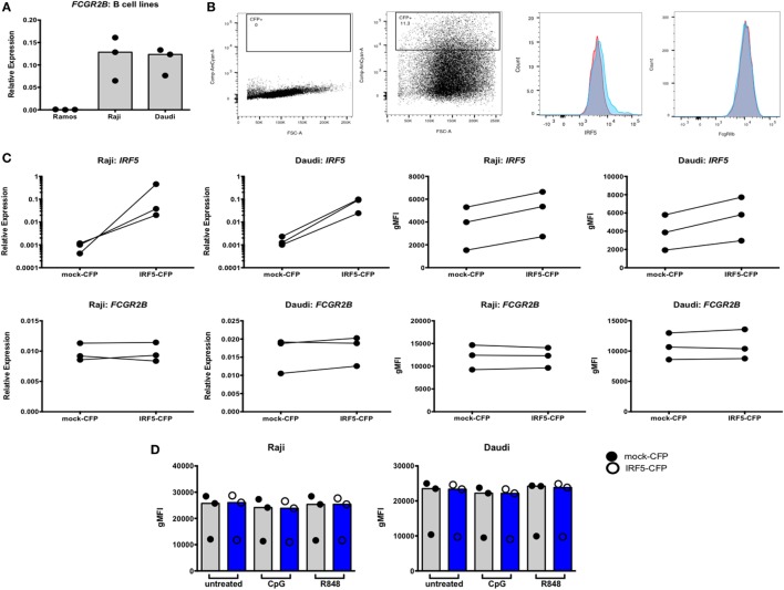
Interferon regulatory factor 5 (IRF5) overexpression does not affect FcγRIIb expression in Raji and Daudi cells. (A) FcγRIIb expression in Ramos, Raji, and Daudi cells. (B) Representative flow cytometric data showing untransfected (far left) and transfected (center left) cells. Gated cells were used for sorting and analyses. Representative histogram of IRF5 protein expression on mock (red) and IRF5 (blue) transfected cells (center right), representative histogram of FcγRIIb protein expression on mock (red) and IRF5 (blue) transfected cells (far right). (C) IRF5 mRNA (top row, far left and left) and protein (top row, far right and right) levels in CFP+ cells. FcγRIIb mRNA (bottom row, far left and left) and protein (bottom row, far right and right) levels in CFP+ cells. (D) FcγRIIb protein levels in CFP+ cells stimulated with CpG or R848 for 24 h. All data shown are representative of three independent experiments.

