Figure 1.
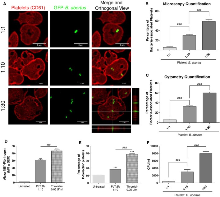
There is a direct interaction between platelets and Brucella abortus. (A) Confocal micrographs of platelets incubated with green fluorescence protein (GFP)-B. abortus at different ratios (Platelet:GFP-B. abortus 1:1, 1:10, and 1:30) for 4 h. Platelet population was stained with an anti-human CD61 primary Ab and Alexa 546-labeled secondary Ab (red). (B) Quantification of platelet–B. abortus interaction by confocal microscopy. The number of platelets counted per experimental group was 200. (C) Quantification of platelet–B. abortus interaction by flow cytometry. Data are expressed as the percentage of platelets associated with B. abortus (GFP-positive platelets) ± SEM of three independent experiments. Platelets were also incubated with B. abortus for 30 min and their activation status was measured as Fibrinogen binding (D) and P-selectin expression (E). The platelet activator thrombin was used as control. Bars represent the arithmetic means ± SEM of three experiments or the percentage of platelets that express P-selectin. MFI, mean fluorescence intensity. (F) Quantification of platelet invasion by B. abortus. Data are expressed as colony-forming units (CFU) per ml. ***P < 0.001 vs. untreated; ###P < 0.001.

