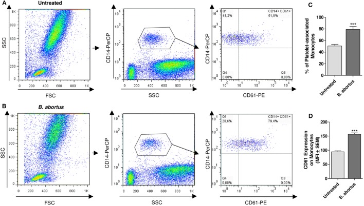
Figure 4.
Brucella abortus infection promotes platelet–monocyte complexes formation in whole blood. Flow cytometry analysis of whole blood untreated (A) or treated with B. abortus (B) for 30 min. Monocyte population was stained with a PerCP-labeled anti-CD14 Ab and platelet population was stained with a PE-labeled anti-CD61 Ab. Cells were plotted on a CD14 vs. SSC dot plot. Then, the CD14+ cells were plotted on a CD14 vs. CD61 dot plot. Finally, the presence of platelet–monocyte complexes (CD14+CD61+) was determined. (C) Quantification of platelet–monocyte complexes within the CD14+ gate. Data are expressed as the percentage of monocytes associated with platelets (CD14+CD61+ cells) ± SEM of three independent experiments. (D) CD61 expression in platelet-bearing monocytes (CD14+CD61+). Bars represent the arithmetic means ± SEM of three experiments. MFI, mean fluorescence intensity. THP-1:PTL:Ba 1:100:100. ***P < 0.001 vs. Untreated.

