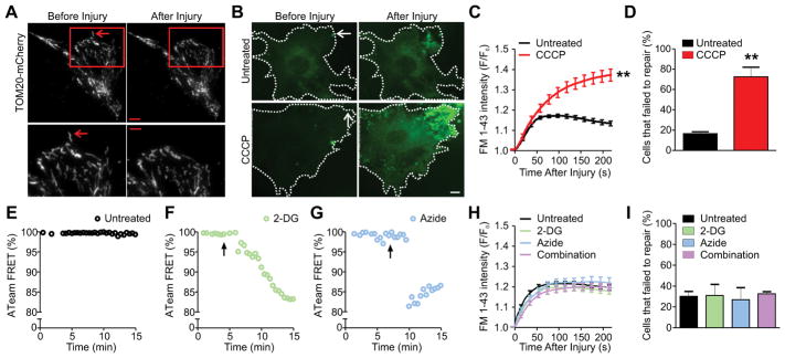
Figure 1. Mitochondria facilitate plasma membrane repair independently of their role in ATP synthesis.
(A) Image of mitochondria localizing near the site of membrane injury by Total Internal Reflection Fluorescence (TIRF) microscopy. Mitochondria are marked by Translocase of the Outer Membrane 20 (TOM20), a structural protein of the outer mitochondrial membrane. Red box (top) indicates magnified areas shown below. Arrows mark the injury site (3 independent experiments). (B and C) Images (B) and quantification (C) of FM 1-43 dye entry in untreated and 5 μM CCCP treated myoblasts (n≥15 myoblasts for each group from 3 independent experiments). Dotted lines mark the cell boundary and arrows mark the injury site. (D) Quantification of myoblasts that failed to repair after laser injury (n≥25 myoblasts for each group from 3 independent experiments). (E–G) Change in YFP/CFP FRET for the cytosolic ATP sensor ATeam monitored in myoblasts that were either untreated (E), treated with 10 mM 2-Deoxyglucose (2-DG) (F), or 10 mM Sodium Azide (Azide) (G). Arrow indicates the time at which drug was added (n≥5 myoblasts for each group from 2 independent experiments). (H) Plot showing kinetics of FM 1-43 dye entry after laser injury of myoblasts pre-treated for 30 minutes (n≥15 myoblasts for each group from 3 independent experiments) with both 10 mM 2-DG and Azide 1 hour before injury. (I) Quantification of myoblasts that failed to repair after laser injury (n≥19 myoblasts for each group from 3 independent experiments). Scale bar=10 μm or 5 μm for magnified image. ** P-value <0.01 by unpaired T-test compared to untreated sample.

