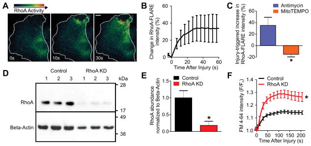
Figure 5. Mitochondrial ROS-dependent RhoA activity is required for repair.
(A and B) Time-lapse images (A) and quantification of change in RhoA-FLARE intensity after laser injury in myoblasts (B) (n=12 myoblasts from 4 independent experiments). (C) Injury-triggered change in the RhoA-FLARE FRET value (RhoA activity) following treatment with 5 μM Antimycin, or 25 μM mitoTEMPO (n=5 or 7 myoblasts respectively from 3 independent experiments). (D) Western blot analysis of RhoA in control or RhoA-targeted miRNA (RhoA KD) treated myoblasts (n=3 independent replicates). (E) RhoA protein abundance in control and RhoA KD myoblasts normalized to loading control (β-actin). (F) Plot showing kinetics of FM 4-64 dye entry after laser injury in control and RhoA KD myoblasts (n≥21 myoblasts for each group from 2 independent experiments). Dotted white line indicates cell boundary. Scale bar=10 μM. *P-value<0.05 by unpaired T-test compared to untreated/control sample.

