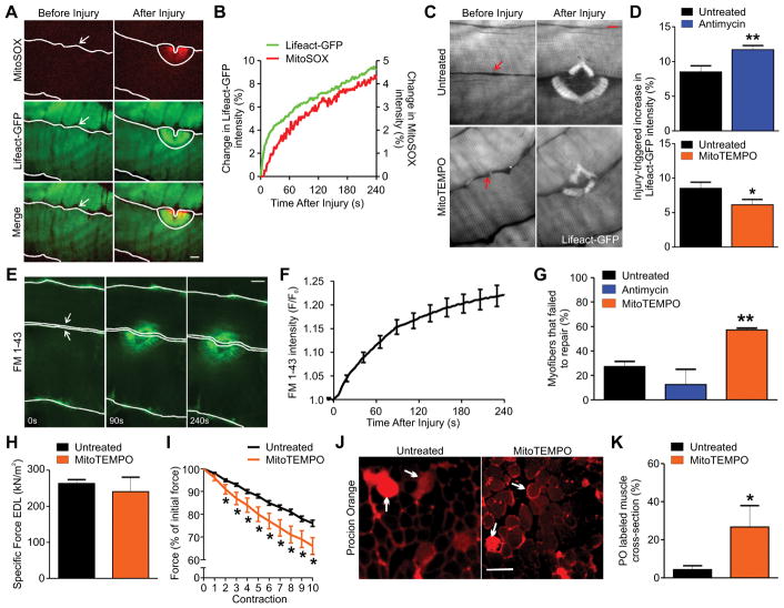
Figure 6. Mitochondrial ROS enables repair of focal and exercise-induced myofiber injury.
(A and B) Image (A) and plot (B) showing mitoSOX and Lifeact-GFP intensity at the injury site for a skeletal myofiber in an intact mouse bicep (2 independent experiments). (C and D) Images (C) and quantification of change in Lifeact-GFP intensity at the injury site in myofibers either untreated or treated with 30 μM Antimycin or 250 μM mitoTEMPO (D) (n≥22 myofibers for each group from 3 mice and 2 independent experiments). (E and F) Images (E) and kinetics of FM 1-43 dye uptake by myofibers after focal laser injury (F) (n=20 myofibers from 3 mice and 2 independent experiments). (G) Plot showing myofibers that failed to repair from focal laser injury. Myofibers were either untreated or treated with 30 μM Antimycin or 250 μM mitoTEMPO (n≥20 myofibers from 2 mice and 2 independent experiments). (H) Specific force of EDL muscles untreated or treated for 20 minutes with 250 μM mitoTEMPO (n=5 mice each from 5 independent experiments). (I) Reduction in EDL muscle contractile force following each round of 20% eccentric exercise (n=5 mice each from 5 independent experiments). (J and K) Images (J) and quantification of Procion Orange (PO) labeling of myofibers in EDL muscle cross section following 10 rounds of eccentric exercise of muscle treated as indicated (K) (n=5 mice each from 5 independent experiments). Arrows mark some of the PO positive fibers. Arrows in laser injury images mark the site of injury and the solid lines mark the myofiber boundary. Scale bar=10 μm (A, C, E) or 100 μm (J). *P-value<0.05 and **P-value<0.01 by unpaired T-test compared to untreated sample or ANOVA followed by Dunnett’s post-test for multiple comparisons.

