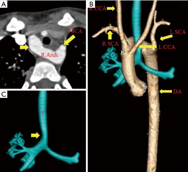
Figure 10.
Right circumflex aorta, a 2-year-old male child with history of chronic cough. CTA (A) demonstrates right aortic arch (short arrow) crossing over to the left side posterior to trachea. After crossing over, the arch gives rise to aberrant left subclavian artery. Volumetric 3D CTA (B) shows right aortic arch with descending aorta on the left side with evidence of tracheal compression by the crossing aortic arch (C). L SCA, left subclavian artery; R arch, right aortic arch; R SCA, right subclavian artery; R CCA, right common carotid artery; L CCA, left common carotid artery; DA, descending aorta; T, trachea.

