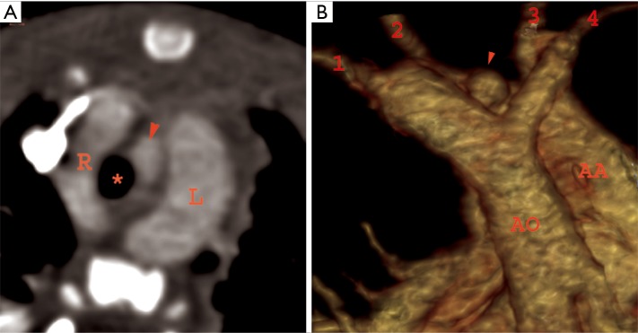
Figure 11.
Double aortic arch: a 15-month-old female with failure to thrive and abnormal mediastinal contour on chest radiograph. Axial CTA image (A) showing double aortic arch encircling the airway (*) with left arch (L) being dominant. Volume rendered image (B) showing four vessel sign in the same patient. Note is made of patent ductus arteriosus (PDA) from left arch (arrowhead). 1, left subclavian artery; 2, left common carotid artery; 3, right common carotid artery; 4, right subclavian artery; R, right; L, left; AO, descending thoracic aorta; AA, ascending aorta.

