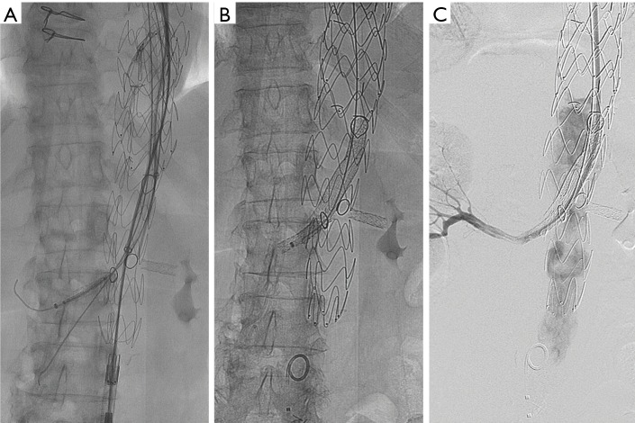
Figure 3.
Seventy yo F with a paravisceral aneurysm that underwent a two stage endovascular repair with a physician modified fenestrated Cook® Zenith® AlphaTM endovascular device. (A) Fluoroscopic image demonstrates successful cannulation of the SMA and right renal artery through the physician created fenestrations. Each fenestration is outlined by a radiopaque marker to allow visualization under fluoroscopy. Of note, the left renal fenestration did not align properly making cannulation of a previously placed left renal iCASTTM (Atrium Medical, Merrimack, NH, USA) stent not possible; (B) scout film shows Gore® Viabahn® stents through the SMA and right renal fenestration creating a branch into their respective target vessels; (C) digital subtraction angiogram of the right renal artery demonstrates adequate flow through the branch. Note the reflux into the aorta demonstrates no significant filling of the left renal artery via the left sided fenestration.

