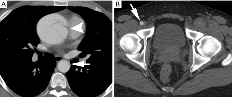
Figure 1.
Contrast-enhanced CT angiography (CTA) of a 29-year-old man with history of Marfan syndrome and hypertension, presenting with severe chest pain radiating to his abdomen with transient paresthesia of left lower extremity. (A) Thoracic imaging confirms an aneurysmal type A aortic dissection originating at the aortic root (arrowhead) and true lumen collapse in descending thoracic aorta (arrow); (B) at the level of the pelvis, note is made of a patent right common femoral artery (arrow) and an occluded left common femoral artery.

