Figure 3.

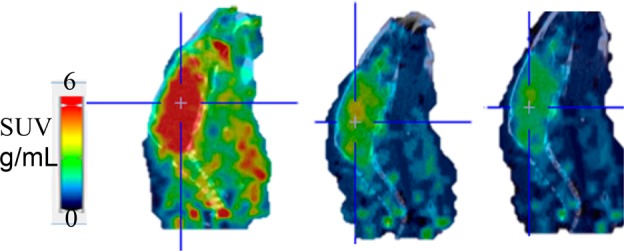
Sum of 0–60 min PET/CT fused sagittal images of [11C]HD-800 in a representative mouse brain (left: baseline; middle: blocking with 5 mg/kg HD-800; right: blocking with 5 mg/kg MPC-6827. Cross lines represent center of brain).

Sum of 0–60 min PET/CT fused sagittal images of [11C]HD-800 in a representative mouse brain (left: baseline; middle: blocking with 5 mg/kg HD-800; right: blocking with 5 mg/kg MPC-6827. Cross lines represent center of brain).