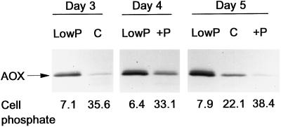
Figure 2.
The level of AOX protein in wild-type tobacco suspension cells grown in complete (C) or low-P medium over a 3- to 5-d period. In some cases, a 3-d-old low-P culture was supplemented with P (+P) and the AOX protein level was examined 1 and 2 d later. For the determination of AOX protein level, washed mitochondria were isolated from cells and the mitochondrial proteins were separated by reducing SDS-PAGE, transferred to nitrocellulose, and probed with a monoclonal antibody to AOX. Numbers along the bottom refer to the total (organic plus Pi) P content of the cells from which mitochondria were isolated. The experiment shown is representative of the trends in AOX protein level observed in several other independent experiments but not always with exactly the same time points being measured.

