
Fig. 2.
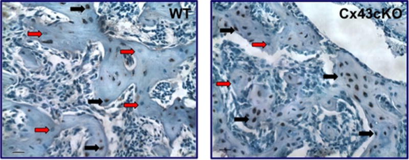
Sclerostin immunohistochemistry. An increase in the number of sclerostin+ osteocytes is observed in the fracture callus of OCN-Cre Cx43-deficient mice relative to WT at 21 days. Black arrows indicate sclerostin+ cells, red arrows indicate sclerostin− cells. Scale bar = 100 microns.