Figure 1.
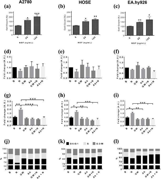
NGF increases the proliferation of A2780, HOSE and EA.hy926 cells.
Cells were stimulated with NGF (25, 50 and 100 ng/ml) for 48 h and then the number of cells was evaluated. (a–c) cell count of A2780, HOSE and EA.hy926 cells after NGF treatment (percentage respect basal condition, n = 3 in triplicate). In subsequent experiments, cells were stimulated with 100 ng/ml NGF, in the presence or absence of the TrkA inhibitor GW441756 (GW; 20 nM) or an NGF-neutralizing antibody (Ab; 5 ug/ml) for 6 h. (d–f) cell death in A2780, HOSE and EAhy.926 cells after NGF treatment (fold change; n = 4); (g–i) semi-quantitative analysis of Ki-67 immunodetection of A2780, HOSE and EA.hy926 cells (fold change, eight images per group, n = 3); (k–m) percentage of cells in different stages of the cell cycle (fold change, n = 4).
Statistically significant changes are indicated as *p < 0.05; **p < 0.01; ***p < 0.001. Statistical analysis, Kruskal–Wallis test.
B, basal condition; HOSE, human ovarian surface epithelial cells; N50, NGF 50 ng/ml; N100, NGF 100 ng/ml; NGF, nerve growth factor; TrkA, tropomyosin receptor kinase A.

