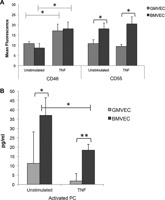
Figure 4.

CD46 and CD55 on the surface of and activated PC in the supernatant of unstimulated and TNF-stimulated BMVECs and GMVECs. A, surface complement regulatory receptors were measured by flow cytometry. BMVECs and GMVECs were incubated with or without TNF (10 ng/ml)-supplemented media for 48 h (without TNF n = 4 for BMVECs and n = 6 for GMVECs; with TNF n = 4 for BMVECs and n = 8 for GMVECs). Samples of 2 × 10⁴ unstimulated or TNF-stimulated BMVECs and GMVECs were labeled with saturating amounts of FITC-conjugated mAbs to CD46 and CD55 or with FITC-conjugated isotype control antibodies. Values are mean fluorescence intensities plus S.D. B, levels of activated PC were measured in BMVEC (n = 4) and GMVEC (n = 5) supernatant after the addition of 0.2 μm PC and 10 nm thrombin over 60 min. The BMVECs and GMVECs in T-75 flasks were either unstimulated or incubated with 10 ng/ml TNF for 48 h before the experiment. *, p < 0.05; **, p < 0.001.
