
Figure 3.
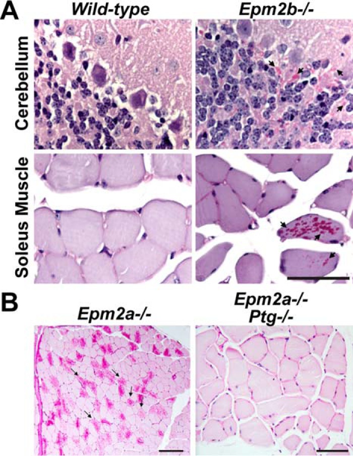
A, periodic acid-Schiff (PAS) staining of cerebellum and soleus sections from WT mice show few stainable structures. Conversely, Epm2b−/− mice exhibit dramatic numbers of stainable polyglucosans, i.e. LBs. Arrows indicate examples of areas rich in LBs. Data are modified from Ref. 32. B, periodic acid-Schiff staining of Epm2a−/− skeletal muscle reveals dramatic accumulations of LBs. Mice lacking both Epm2a and Ptg display minimal to no LBs. Data are modified from Ref. 89.