Figure 1.

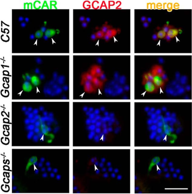
GCAP2 is expressed in the mouse cones. Dissociated retinal cells from mice of the indicated genotypes were incubated with the mCAR antibody (green) to label cones followed by incubation with GCAP2 antibody (red). Arrowheads point to the positions of the cones in each field. Nuclei were stained with DAPI (blue). Images shown are representative of 38 cones from 14 fields (C57), 26 cones from 10 fields (Gcap1−/−), 10 cones from seven fields (Gcap2−/−), and nine cones from three fields (Gcaps−/−). Scale bar, 20 μm.
