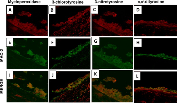
Figure 6.

Macrophages, myeloperoxidase, and myeloperoxidase products co-localize in the arterial wall of 5/6 nephrectomized mice on high-fat, high-cholesterol diet. Representative immunofluorescence and double labeling in the aortic cross-sections from male LDLr−/− 5/6 nephrectomized mice on 24 weeks of high-fat, high-cholesterol diet for Mac-2 (macrophage marker), myeloperoxidase, 3-chlorotyrosine, 3-nitrotyrosine, and o,o′-dityrosine are shown. Shown is a section view of the aortic wall double-labeled for Mac-2 (green; E–H) and MPO (red, A), 3-chlorotyrosine (red, B), 3-nitrotyrosine (red, C), and o,o′-dityrosine (red, D). In all sections, the signals of MPO, o,o′-dityrosine, 3-nitrotyrosine, and 3-chlorotyrosine co-localized with Mac-2 (yellow, I–L).
